Spinal Anatomy & Glossary
Spine Anatomy
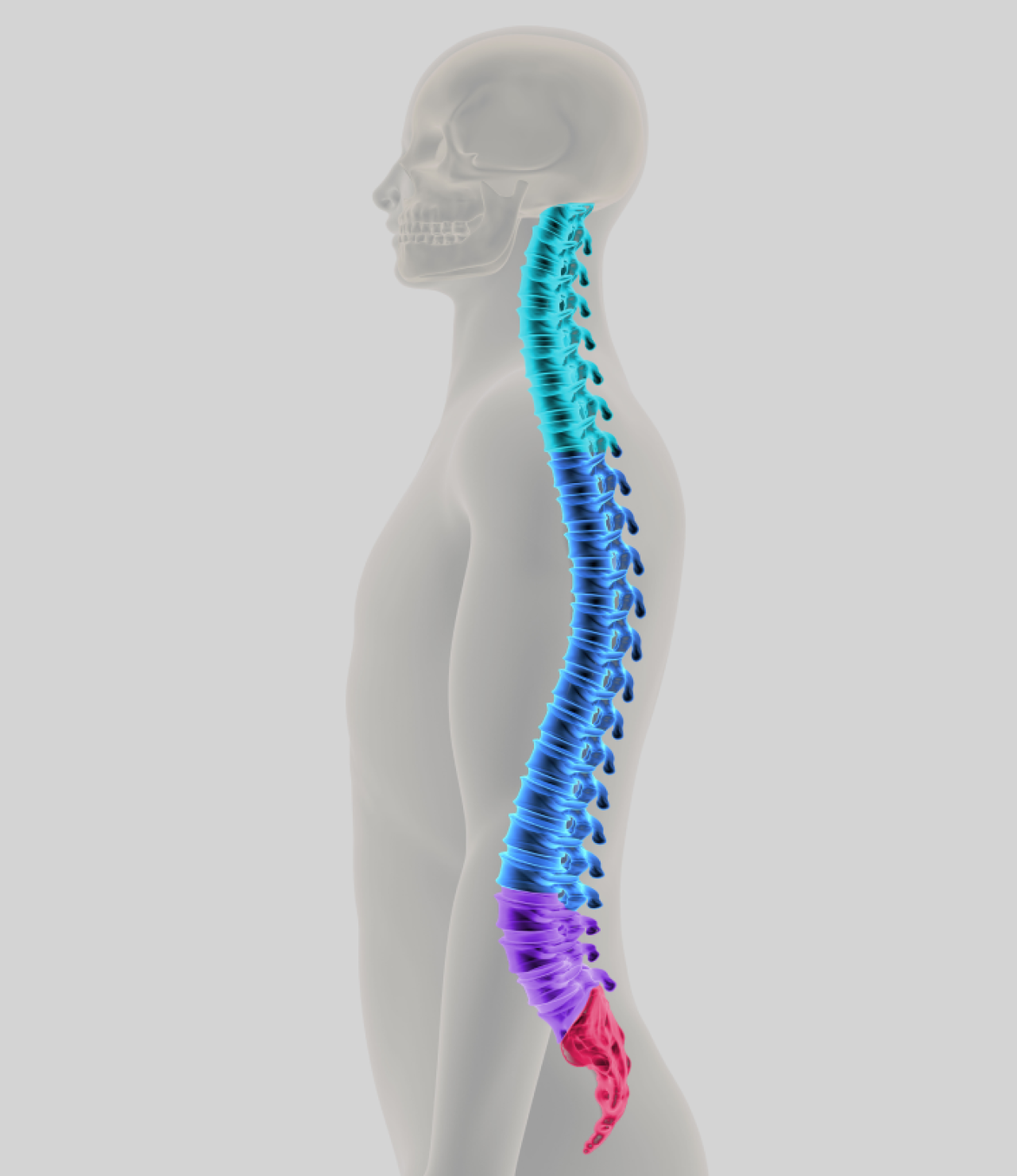
Made up of bony segments called vertebrae and fibrous tissue called intervertebral discs, the spine (back bone) gives you stability and smooth movement. It also protects your delicate spinal cord.
The vertebrae and discs form a column from head to pelvis and can be divided into four parts:
Spine Anatomy
Made up of bony segments called vertebrae and fibrous tissue called intervertebral discs, the spine (back bone) gives you stability and smooth movement. It also protects your delicate spinal cord.
The vertebrae and discs form a column from head to pelvis and can be divided into four parts:
1
Cervical region [neck]
At the very top, made up of seven small vertebrae.
2
Thoracic region
[mid-back]
n the middle, made up of 12 vertebrae from which the ribs are hinged
3
Lumbar [lower back]
these vertebrae are the largest of the mobile vertebra and support two thirds of your body’s weight
4
Sacrum (between your hips) and coccyx (tail bone)
in the lowest region of the spine, the sacrum is a triangular plate made up of five fused vertebral segments and the four coccyges make up what is commonly called the tail bone.
Spine Glossary
Adolescent scoliosis
Lateral spinal curvature that appears before the onset of puberty and before skeletal maturity
Adult scoliosis
Curvature of the spine of any cause, which is present after skeletal maturity
Ankylosing spondylitis
Often called ‘bamboo spine disease’, this inflammatory disease of the spine gradually restricts movement and is most common in young adults resulting in morning pain
Anterior longitudinal ligament
A ligament that attaches to the anterior (front) of every vertebra, from the base of the skull to the sacrum (the area of spine between your hips)
Anterior sacroiliac ligaments
Ligaments that span the front of the sacroiliac joints (which connect the hip bone to your spine), from the sacrum to your hip area. They are not as strong as those along the posterior (back)
Anterior spinal fusion
A surgical technique that involves replacing the disc between two vertebrae with a bone graft. Additional structural supports may be placed in the disc space, such as hard (cortical) bone grafts, metal or synthetic spacers
Anterior superior iliac spine
This is what you may think of as your hip bones – they jut out each side of your pelvis
Anterior
The front portion of the vertebral body. It may also indicate the position of one structure relative to another
Anteroposterior view (AP view)
An X-ray in which the patient faces toward the X-ray beam, which passes from anterior to posterior (front to back) through the patient, and away from the X-ray film
Apex of scoliosis
The area of greatest curvature or displacement from the midline of the body
Apical vertebra
When referring to scoliosis, it is the vertebra with the greatest distance from the midline and has the most rotation.
Apophysis
A growth plate that is not apparent on X-rays until the bone is maturing, when it begins to ossify (harden). The iliac apophysis is often used to estimate a child’s skeletal maturity
Arachnoid
A thin layer of connective tissue surrounding the brain that contains the spinal fluid around the spinal cord.
Artery of adamkiewicz
An artery that supplies blood to the front of the spine. The level at which it enters the spinal canal varies widely, and if the blood flow in this artery is impaired it can cause symptoms that range from weakness to full paralysis
Ataxic gait
Uncoordinated, unsteady walking with the feet spread apart (wide-based) which may be caused by either spinal stenosis (narrowing of the spaces within your spine) or a brain (central nervous system) disease
Atlanto-axial instability
Abnormal, excessive motion between the first and second cervical (neck) vertebrae, which may be due to disease or injury
Atlanto-axial rotatory subluxation
When the first and second cervical (neck) vertebrae are out of position, causing the head to tilt to one side, and can’t turn to the other side. This can be hard to diagnosis and may require a CT scan.
Atlanto-dens interval
A measurement taken from the side of the neck. This looks at the space in between the front ring of the first vertebra and the front of a bony protrusion on the next. Increased space can indicate damage to the supporting ligaments caused by illness or injury
Atlanto-occipital dislocation
A separation of the upper cervical spine from the skull after injury. This is often fatal.
Atlanto-occipital joint
The joint between the skull and the first cervical (neck) vertebra (c1)
Atlas
The first cervical (neck) vertebra (c1) to which the skull attaches above and the axis (c2) attaches below
Autologous blood
Blood collected from a person for later transfusion to that same person. This technique is often used before elective surgery
Autonomic dysreflexia
This involves over-stimulation of the autonomic nervous system in a paraplegic patient whose spinal injury is at or above t6 (the vertebrae in the mid back). This can create excessive sweating, goose flesh, headaches, high blood pressure, slow heart rate and unusual flushing of the skin. It can be caused by infection, over-distention of the urinary bladder, constipation or a skin wound. If untreated, it can become a medical emergency or cause death
Autotransfusion
Transfusing previously drawn blood back to the same patient.
Axis
The second cervical (neck) vertebra made up of a ring of bone and the odontoid, a bony protrusion. As it moves with the atlas (c1), it provides 50% of the cervical spine rotation
Basilar impression/invagination
When the upper cervical (neck) spine presses into the base of the foramen magnum (skull), the spinal cord or brainstem can be compressed. Diseases that soften bones may cause this.
Bilateral facet dislocation
Usually caused by severe injury to both sides of the neck and its soft tissue. This lets one part of the vertebra move forward on the one below and requires surgery.
Biomechanical back pain
Brought on by muscular strain/ligamentous injury, this causes other muscles or structures to become stressed and painful.
Block vertebrae
When a person is born with two or more fused vertebrae
BMP
Bone morphogenetic protein is a genetically engineered protein that stimulates bone production to help them heal and/or fuse. These proteins are made by our bodies, but in much smaller quantities. BMP is added to your bone but is not yet FDA-approved for all types of surgery
Body cast
A cast that surrounds the chest, abdomen and pelvis and may also include the shoulders. This may be used to correct scoliosis in very young patients or to keep patients still after surgery.
Bone graft
Human bone, harvested from one part of the body, and grafted to another. The hip bones are a common place to take bone grafts.
Bone spur
An overgrowth of bone in response to stress or injury.
Cervical spine
Seven spinal segments (c1-c7) between the base of the skull (occiput) and the thoracic spine. The normal cervical spine alignment is called lordosis.
Cervicothoracolumbosacral orthosis (CTLSO)
A type of brace that keeps the spine still from the neck to the lumbar. This may be used to help treat or limit the progress of scoliosis curve(s) while a child is growing, or to keep the spine still after surgery.
Coccyx
Also called the tailbone, this is the lowest segment of the spine.
Compensatory curve
This is a curve above or below a deformity, which develops to maintain normal body alignment
Congenital scoliosis
Scoliosis (curved spine) caused by bony abnormalities of the spine present at birth.
Corpectomy
The surgical removal of all or part of the vertebral body.
Decompensation
In scoliosis (curved spine), this refers to loss of spinal balance when the thoracic (rib) cage is not centred over the pelvis
Decompression
This treatment aims to relieve pressure on the spinal cord or nerve roots caused by bone, bone fragments, disc herniation (slipped disc), ligaments, bone spurs, tumour, infection or abnormal curvature of the spine (scoliosis or kyphosis)
Disc degeneration
The loss of the fluid content, structure and functional integrity of the disc
Disc herniation
Commonly called a slipped disc, this is when the fibrous tissue of a spinal disc is torn, causing it to slip and cause a bulge
Discectomy
Removal of all or part of an intervertebral disc (the soft tissue that acts as a shock absorber between the vertebral bodies). This may be done for fusion or to fix herniation
Distal
Situated away from or farther from a point of reference; opposite of proximal
Double curve
Two lateral curvatures (scoliosis) in the same spine
Double major curve
Describes two structural spinal curves, usually of equal size
Double thoracic curve
Describes a structural curve in the upper part of the mid back (thoracic), a larger, more deforming curve in the lower part and a less serious curve in the lumbar
Dura
The three-layer membrane that contains the spinal cord and spinal fluid. It can be torn or stretched during spinal surgery, and can often be repaired or patched
Gardner-Wells tongs
A device used to position the head or apply traction to the neck during surgery. The tongs are attached to the skull with a screw above each ear after the patient is asleep
Hemivertebra
An abnormality of a vertebral body caused when one side of a vertebra doesn’t develop completely before birth. This is usually a wedge shape, which causes scoliosis or kyphosis
Hyperkyphosis
Kyphosis refers to an abnormal increase in the forward curvature
Hysterical scoliosis
A non-structural deformity of the spine caused in response to a psychological disorder.
Idiopathic scoliosis
A curve in the spine for which the cause has not been established
Iliac bone
A part of the pelvic bone that is above the hip joint and from which autogenous bone grafts are frequently obtained
Inclinometer
An instrument used to measure the angle of thoracic (rib) or lumbar (flank) prominence, referred to as the angle of trunk rotation (ATR)
Infantile scoliosis
A curvature of the spine that develops before the age of three
Internal fixation
The immobilisation of bone fragments or joints with implants (metal screws, rods, etc.) to promote healing or fusion
Interspinal or intervertebral disc
The structure that normally occupies the space between two moving vertebrae, more prominent in the neck and lower back. The centremost portion of the disc is normally made up of a clear gelatinous material that varies in consistency from jellied to very thick and less pliable.
Juvenile scoliosis
Scoliosis (curve of the spine) developing between the ages of three and ten years.
Kyphoscoliosis
A structural scoliosis (curve of the spine) associated with increased kyphosis (round back).
Kyphosis
The normal forward curvature of the thoracic (mid-back) spine.
Lamina
Part of a vertebral arch that makes up the roof of the spinal canal, through which runs the spinal cord
Laminectomy
An operation to remove part or all of the lamina to minimise an intervertebral disc protrusion or to decompress a nerve root.
Lateral
Situated away from the midline of the body.
Lordoscoliosis
A lateral curve in the spine associated with increased lordosis (swayback)
Lordosis
The normal inward curve of the lumbar spine (just above the buttocks)
Lumbar curve
Also known as lumbar scoliosis, this is a curve in the spine between the first and fourth lumbar (lower back) vertebrae
Lumbar spine
Five mobile segments of the lower back (l1 to l5). These are the largest of the vertebral segments and provide most of the bending and turning ability of the back. They also bear most of the weight of the body
Lumbosacral curve
Also known as lumbosacral scoliosis, this is when the spine curves to the side, at the fifth lumbar vertebra or below
Lumbosacral
Pertaining to the lumbar and sacral regions of the back
Medial
Situated closer to the midline of the body
Nerve root
The portion of a spinal nerve close to its origin from the spinal cord
Osteotomy
Surgery to remove a wedge or piece of vertebral bone to alter the alignment of the spine. This may also be used in previously fused vertebrae to enable the surgeon to move them
Pedicle
The part of each side of the neural arch of a vertebra that projects backward from the vertebral body. It connects the lamina with the vertebral body
PLIF – posterior lumbar interbody fusion
A technique for performing an anterior fusion (front of the spine) of two vertebrae from a posterior (behind) approach
Posterior longitudinal ligament
A ligament that attaches to the back of every vertebra, from the base of the skull to the sacrum
Posterior spinal fusion
A surgical technique to roughen or remove hard bony surfaces of the lamina(e), back of the vertebrae and facet joints (between the vertebrae in the back of the spine). This stimulates two or more vertebrae to fuse and may be combined with a bone graft or implant
Posterior
Located behind a structure, and relating to the back side of the body
Primary curve
The first, or earliest, curve to appear
Proximal
Nearer or closer to a point of reference; opposite of distal
Pseudarthrosis
An area of the spinal fusion where the bone did not fuse (heal), often found with broken instrumentation and, in some instances, increased pain
Risser sign
Used to evaluate skeletal and spinal maturity, this refers to a crescent-shaped line of bone that appears across the top of each side of the pelvis
Sacral spine – (sacrum)
The curved triangular bone at the base of the spine, consisting of five fused segments of the lower spine. The sacrum articulates (connects) with the last lumbar vertebra and the pelvic bones
Sacroiliac joint
The joint between the ilium (upper parts of the hip) and sacrum (the spine between the hip bones), one each side of the pelvis. This joint has a small amount of motion and may be a source of low back pain
Sacroiliitis
Inflammation within the sacroiliac joint, which is beween the upper parts of the hips and the sacrum (the spine between the hip bones). This may be an associated symptom of ankylosing spondylitis
Sciatica
A common term to describe pain along a sciatic nerve, especially noted in the back of the thigh and below the knee
Scoliometer
A name for an inclinometer used to measure trunk rotation
Scoliosis
An abnormal curve of the spine. Rotation of the vertebrae also produces rib cage and flank muscle
Spinal canal
The long canal through which the spinal cord passes from the brain down to the lumbar segment in adults. Below this level are numerous nerve roots from the spinal cord that resemble a horse’s tail. The thick outer covering of the spinal cord is called the dura
Spinal fusion
A surgical procedure to permanently fuse two or more vertebrae with a bone graft. It can be done from the anterior (front), posterior (back) or as a staged procedure (first anterior and then posterior), usually with metal implants
Spinal instrumentation
Metal implants fixed to the spine to improve spinal deformity while the fusion becomes solid bone. This includes a wide variety of rods, hooks, wires and screws
Spinal stenosis
Narrowing or reduction in the diameter of the spinal canal which surrounds the spinal cord, spinal roots and spinal sac (dura). This may be caused by disc herniation, bone fracture, arthritic overgrowth of bone and soft tissue, or occasionally may have developed before birth
Spinous processes
The portion of the vertebrae that stick out to create the bumps felt on the midline of the back
Spondylitis
An inflammatory disease of the spine
Spondylolisthesis
A slipping of a vertebra onto the lower vertebra. There are several causes for this and can vary in severity
Spondylolysis – (also referred to as a stress fracture or a pars fracture)
A stress fracture in the lower back through the pars interarticularis, a thin bone segment joining two vertebrae
Structural curve
A segment of the spine that has fixed (nonflexible) lateral curvature
Thoracic (dorsal) spine
Twelve spinal segments (t1-t12) incorporating the ribs. Other than a slight increase in size from top to bottom, they are fairly uniform
Thoracic curvature
Any spinal curvature in which the top of curve is between the second and eleventh thoracic vertebrae (mid back)
Thoracolumbar curve
Any spinal curvature in which the top of curve is at the twelfth thoracic or first lumbar vertebra (mid-to-lower back)
Thoracolumbosacral orthosis (TLSO)
A type of brace to keep the thoracic lumbar and sacral spine still (the part of the spine from your lower back to your bottom). This may be used to help stabilise or prevent a scoliosis curve from progressing while a child is growing, or to keep the spine still after surgery
TLIF – transforaminal lumbar interbody fusion
A minimally invasive technique for performing an anterior fusion (front of the spine) of two vertebrae from behind
Transition syndrome
A degenerative change with bony instability above or below a previous fusion
Vertebra
One of the 33 bones that make up the spinal column
Vertebral column
The flexible supporting column of vertebrae separated by discs and bound together by ligaments
Useful links